
Cart

Oops, your cart is still empty
Looks like you haven’t added anything yet. Start exploring our products and find what you need.
Research use only. The compounds referenced on this page are research-grade reference materials for laboratory use only. They are not for human or veterinary use.
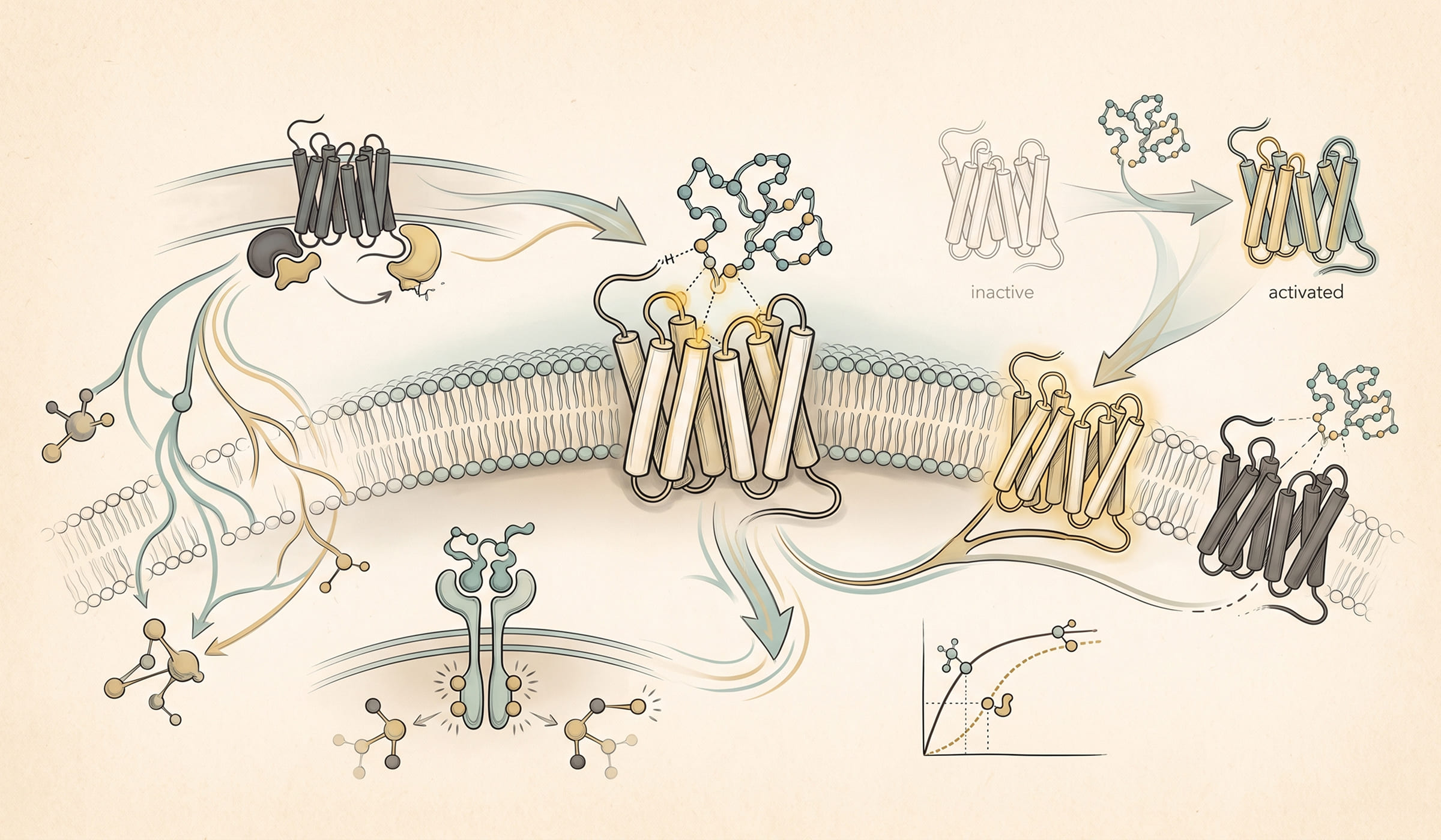
Peptide-receptor interactions, governed by binding affinity, selectivity, and the mechanism of receptor activation, determine the pharmacological effect observed in research models. Understanding the major receptor classes and the binding metrics that characterize peptide-receptor interactions is essential for interpreting peptide research literature and reasoning about mechanism of action claims.
This article covers the receptor families most relevant to research peptides, the affinity metrics that quantify binding, the binding mechanisms (competitive, allosteric, non-competitive), and the selectivity considerations that shape modern analog design.
Peptide ligands act at several receptor classes and the two that dominate this literature behave differently. For the general chemistry these molecules share, see what peptides are and how they are made. The table separates them so the research readouts in the sections below can be read against the right mechanism.
| G protein-coupled receptors | Receptor tyrosine kinases | |
|---|---|---|
| Signal transduction | Conformational change couples the receptor to heterotrimeric G proteins and to arrestins. | Ligand binding drives receptor dimerization and autophosphorylation of intracellular tyrosine residues. |
| Timescale | Seconds to minutes for second-messenger responses. | Minutes to hours for the phosphorylation cascades and transcriptional responses. |
| Typical peptide ligands | Melanocortin peptides, opioid peptides, many neuropeptides and gut peptides. | Growth factor peptides and proteins acting at kinase receptors. |
| Common research readouts | Second messenger accumulation, arrestin recruitment, receptor internalization, binding kinetics. | Receptor phosphorylation, downstream kinase activation, transcriptional readouts. |
| Where selectivity comes from | Receptor subtype differences and, increasingly, biased signaling toward particular transducers. | Receptor family membership and the specific downstream adaptors recruited. |
Biased agonism is the concept doing most of the current work in the GPCR literature: different ligands at the same receptor can favor different transducers rather than simply producing more or less of one response. Recent structural and pharmacological studies have examined how that bias arises (Nat Commun, 2024; PMID 38824166; Nature, 2023; PMID 37532940), and receptor kinase specificity has been shown to shape which ligands produce it (Commun Biol, 2024; PMID 38956302). Melanocortin-derived peptides are one worked case on this site, covered in KPV tripeptide and NF-kB signaling. Compounds without an established receptor are handled differently, as in BPC-157 mechanism of action research.
Most peptide receptors fall into one of four major families.
GPCRs are integral membrane proteins with seven transmembrane alpha helices. The N-terminus is on the extracellular side and the C-terminus on the intracellular side. The extracellular regions and the upper portions of the transmembrane helices form the ligand binding site for many peptide GPCRs, while the intracellular regions couple to heterotrimeric G proteins.
GPCRs are the most abundant single class of cell surface receptors and are engaged by a remarkable diversity of ligands, including small molecules, lipids, photons (in rhodopsin), and peptides. The peptide GPCRs of greatest relevance to research peptide work include:
GPCR signaling is mediated by G proteins (Gs, Gi, Gq, G12/13) that activate or inhibit second messenger systems including cAMP, calcium, and inositol phosphates.
Receptor tyrosine kinases are single-transmembrane proteins with an extracellular ligand binding domain and an intracellular tyrosine kinase domain. Ligand binding induces receptor dimerization, kinase activation, and downstream phosphorylation cascades that ultimately regulate gene expression and cell behavior.
The IGF-1 receptor and the related insulin receptor are the most relevant RTKs for research peptide work. IGF-1 LR3 (a long arginine 3 analog of IGF-1) engages the IGF-1 receptor and has a substantial preclinical literature.
Ion channels mediate fast electrical signaling at synapses and across cell membranes more broadly. Some peptides engage ion channels directly. Conotoxins, derived from cone snail venoms, are among the most studied peptide ion channel modulators in research and have provided high-affinity selective probes for many ion channel subtypes (Lewis et al., 2012; PMID 22383759).
Nuclear receptors are intracellular ligand-activated transcription factors. Most engage small molecule ligands rather than peptides, but a few peptide ligands are known. Peptide engagement of nuclear receptors is comparatively rare in research peptide work.
Quantitative pharmacology rests on standardized affinity and potency metrics.
The dissociation constant Kd is the equilibrium concentration of free ligand at which half of the receptors are bound. It is the most direct measure of binding affinity. A Kd of 1 nM is high affinity; a Kd of 1 micromolar is moderate to weak.
Kd is determined experimentally by saturation binding (varying the concentration of labeled ligand and measuring receptor occupancy) or by competition binding (using a fixed concentration of labeled ligand and varying concentrations of an unlabeled competitor). Surface plasmon resonance (SPR), bio-layer interferometry, and fluorescence anisotropy are common biophysical methods for Kd determination.
EC50 is the concentration at which a peptide produces 50 percent of its maximal functional response in a cellular assay. The assay readout might be cAMP, calcium flux, gene reporter activity, or any downstream functional measure. EC50 reflects both binding affinity and intrinsic activity (efficacy).
EC50 is not necessarily equal to Kd. For systems with receptor reserve (more receptors than needed for maximum response), EC50 can be substantially lower than Kd, because half-maximal response is achieved at low receptor occupancy.
IC50 is the concentration of an inhibitor that produces 50 percent inhibition of a measured response. It depends on the concentration of competing agonist or substrate in the assay and is therefore an apparent value. The Cheng-Prusoff equation converts IC50 to Ki (the equilibrium inhibition constant) when the competing agonist’s Km or Kd is known.
In the modern literature, affinity values are often reported as their negative base-10 logarithms. pKd 9 corresponds to Kd of 1 nM; pKd 6 corresponds to Kd of 1 micromolar. The logarithmic scale facilitates comparison across orders of magnitude in affinity.
Read more: Peptide Structure-Activity Relationships (SAR): Principles for Researchers
GPCRs are the most common research peptide targets, so understanding how peptides engage GPCRs deserves special attention.
Modern structural biology has produced atomic-resolution structures of many GPCRs in agonist-bound and antagonist-bound forms. For peptide GPCRs, the ligand binding site typically extends from the extracellular vestibule into the upper part of the transmembrane bundle. Long peptides can engage extracellular surfaces extensively; short peptides typically reach into the transmembrane core.
Receptor activation involves outward movement of transmembrane helix 6 on the intracellular side, which opens a cavity for G-protein binding. The energy of ligand binding propagates through the receptor to drive this conformational change.
An agonist stabilizes the active receptor conformation and triggers G-protein binding and downstream signaling. An antagonist binds without stabilizing the active conformation, blocking endogenous agonists from binding. A partial agonist stabilizes an intermediate conformation that produces less than maximal signaling. An inverse agonist stabilizes an inactive conformation and reduces constitutive (basal) signaling.
The choice between agonism and antagonism is rarely made in isolation; modern research peptide design considers receptor subtype selectivity, biased agonism (preferential activation of one downstream pathway over another), and pharmacokinetic factors as well.
Receptor families often contain multiple subtypes with overlapping but distinct pharmacology. The melanocortin receptor family includes MC1R, MC2R, MC3R, MC4R, and MC5R, each with different physiological roles. The opioid receptor family includes mu, delta, kappa, and nociceptin receptors. Selectivity for one subtype over another can dramatically alter the observed pharmacology and is often a key research design objective.
Selectivity arises from sequence and structural differences between subtypes that produce different binding pocket geometries. Peptide analog design programs systematically modify residues that contact the variable regions of the binding pocket to tune selectivity.
Read more: Peptide Classification Systems: Linear, Cyclic, Branched, and Stapled Peptides
Binding parameters in this literature come from a small number of established approaches, and knowing which was used matters when comparing values across papers. Isothermal titration calorimetry measures binding thermodynamics directly in solution (Methods Mol Biol, 2024; PMID 38019443). Real-time kinetic methods using immobilized receptor resolve association and dissociation rates rather than an equilibrium constant alone (Anal Chem, 2026; PMID 42423075). Structural methods establish the binding mode itself, as in recent work on ligand recognition and G protein coupling at a peptide-responsive receptor (Nat Commun, 2023; PMID 37591889).
An affinity value without its method and conditions is not comparable to another, which is the same principle that applies to purity figures on a certificate of analysis. Pathway-level literature without a defined receptor is covered in GHK-Cu wound healing pathway research and summarized in the healing peptides research overview. Compound-level data is indexed under chemical reference data by compound and the stocked material in the research peptide catalog.
Several binding modes have been characterized for peptide-receptor interactions.
In competitive binding, the ligand and a competing molecule (often the endogenous agonist) bind to the same site, producing mutually exclusive occupancy. Competitive antagonists shift the agonist concentration-response curve to the right (higher concentrations needed to achieve the same effect) without reducing the maximum response.
Allosteric modulators bind to a site distinct from the orthosteric (canonical) ligand binding site. Positive allosteric modulators (PAMs) increase the affinity or efficacy of the orthosteric agonist; negative allosteric modulators (NAMs) decrease them. Allosteric modulation often shows ceiling effects (the maximum modulation is bounded), which can be advantageous for fine-tuning physiological responses.
Peptide allosteric modulators are emerging as a research direction, particularly for GPCRs where small-molecule allosteric modulators have already been validated. The receptor selectivity advantages of allosteric modulation can complement orthosteric peptide agonists in combination research strategies.
Some antagonists bind so tightly or so persistently that increasing agonist concentration cannot fully overcome their effect. This produces a depression of the maximum response in addition to the rightward shift seen with classical competitive antagonism. Irreversible antagonists, including covalent binders, fall into this category.
Selectivity is one of the most important considerations in peptide research design.
Selectivity arises from sequence differences in the receptor binding pocket between subtypes. Modern analog design uses these differences to engineer subtype selectivity. The dual GIP and GLP-1 agonism of tirzepatide is a deliberate design choice that exploits structural similarities between the two related receptors (Coskun et al., 2018; PMID 30404862). The triple agonism of retatrutide extends this approach (Jastreboff et al., 2023; PMID 37296781).
A peptide that engages an unintended receptor in an experimental system can confound the observed pharmacology. Selectivity profiling on a panel of related and unrelated receptors is standard practice for novel research peptides. Selectivity ratios (the ratio of EC50 at the target receptor to EC50 at off-target receptors) are reported in well-conducted SAR papers.
Commercial receptor panel screening services test compounds against dozens or hundreds of receptors and report binding or functional activity. Standard panels include the GPCR panel from Cerep (Eurofins), the kinase panel from Reaction Biology, and various ion channel panels. For research peptides, GPCR panel screening is typically the most informative.
Modern pharmacology has moved beyond simplified models, and the modern conceptual framework is worth understanding.
The key-lock metaphor introduced by Emil Fischer in 1894 framed receptor pharmacology for a century. The metaphor envisions the ligand and the receptor as rigid complementary shapes, with binding requiring exact geometric fit.
The key-lock model captures the importance of complementary shape and chemistry in receptor recognition, and it remains a useful pedagogical tool. Modern structural biology has shown that the metaphor is incomplete in important ways.
The induced fit model, introduced by Daniel Koshland in 1958, recognized that the receptor adapts its shape upon ligand binding. The receptor and ligand adjust to one another, producing the bound state through a coupled conformational change. Induced fit explains many observations that the rigid key-lock model cannot, including the dependence of binding affinity on protein flexibility and the existence of multiple receptor conformations.
The most modern framework, conformational selection, recognizes that receptors exist in equilibrium between multiple conformations even in the absence of ligand. The ligand selects (preferentially binds) one of the pre-existing conformations, shifting the equilibrium. This framework integrates allosteric communication, biased signaling, and the dependence of pharmacology on receptor expression context. It has become the dominant conceptual framework in modern receptor pharmacology research.
Read more: What Are Peptides? A Comprehensive Research Reference Guide
Kd is the equilibrium dissociation constant; it measures binding affinity at the molecular level (the concentration at which half the receptors are occupied). EC50 is the concentration that produces half-maximal functional response in a cellular assay. The two often differ because of receptor reserve, downstream amplification, and assay-specific factors. Kd is the more fundamental molecular property; EC50 is the more practical functional readout.
A low Kd is favorable but not sufficient. Selectivity for the intended target over other receptors, metabolic stability, cell penetration (for intracellular targets), and pharmacokinetic properties all contribute to whether a peptide is a useful research tool or a viable drug candidate. A peptide with Kd of 100 nM and excellent selectivity may be more useful than a peptide with Kd of 1 nM and poor specificity.
Allosteric modulators bind to sites distinct from the orthosteric (canonical) ligand pocket and modulate the affinity or efficacy of the natural ligand. Positive allosteric modulators enhance natural signaling; negative allosteric modulators reduce it. Allosteric peptides offer selectivity advantages, ceiling effects on activity, and complementary mechanisms that may have fewer off-target effects than orthosteric agonists.
Common methods include surface plasmon resonance (SPR), bio-layer interferometry, fluorescence polarization, radioligand displacement assays, and cellular functional assays followed by Cheng-Prusoff conversion. Each method has trade-offs in sensitivity, throughput, and the kinetic information obtained. SPR provides binding kinetics (on-rate, off-rate) in addition to equilibrium affinity.
Yes. Some peptides are designed for multi-receptor activity (the dual GIP and GLP-1 agonism of tirzepatide, the triple agonism of retatrutide). Others activate multiple receptors as an unintended off-target effect. Both cases are studied carefully in research analog design, with selectivity panels guiding which co-activations are desirable and which are undesirable.
A G protein-coupled receptor transduces ligand binding through a conformational change that couples it to G proteins and arrestins, on a timescale of seconds to minutes. A receptor tyrosine kinase responds to ligand binding by dimerizing and autophosphorylating intracellular tyrosine residues, driving kinase cascades over minutes to hours.
Biased agonism describes ligands at the same receptor favoring different downstream transducers rather than producing more or less of a single response. It is a central concept in current GPCR pharmacology because it means two ligands with similar affinity can produce different signaling profiles.
Isothermal titration calorimetry measures binding thermodynamics directly in solution. Real-time kinetic methods using immobilized receptor resolve association and dissociation rates. Structural methods establish the binding mode itself. An affinity value is only comparable to another measured by a comparable method under comparable conditions.
Research-only disclaimer. The peptides described in this article are sold and discussed for laboratory and research purposes only. They are not intended for human consumption, diagnostic use, or therapeutic application.
Educational notice. This article is for educational and informational purposes only and is intended for licensed researchers and laboratory professionals. The peptides discussed are research chemicals sold for laboratory and research applications. They are not intended for human consumption, diagnostic use, or therapeutic application.


